
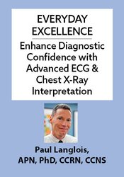
Everyday Excellence: Enhance Diagnostic Confidence with Advanced ECG & Chest X-Ray Interpretation
- Speaker:
- Paul Langlois, APN, PhD, CCRN, CCNS
- Duration:
- 6 Hours 20 Minutes
- Format:
- Audio and Video
- Copyright:
-
Nov 05, 2025
- Product Code:
- POS078120
- Media Type:
- Digital Seminar

Description
- Master high-level 12-lead ECG patterns across a range of clinical scenarios
- Mitigate chest x-ray errors with advanced interpretation techniques
- Integrate evidence-based diagnostic guidelines
- Sharpen clinical decision making and expand professional autonomy
Whether you’re working in primary care, urgent care, or acute settings, NPs, PAs, and RNs play a pivotal role in early recognition and management of critical conditions. Mastering diagnostic tools like the 12-lead ECG and chest x-ray is essential for accurate assessment, timely intervention, and improved patient outcomes.
Even experienced clinicians can find interpretation of these tools challenging. This high-impact, evidence-based one-day seminar will provide you with practical, actionable skills through real-world patient case studies and current best practice guidelines.
Key Findings You’ll Learn:
12-Lead ECG Interpretation
- Myocardial infarction and ischemia
- Arrhythmias
- Bundle branch blocks
- Electrolyte disturbances
- Pericarditis and brugada syndrome
- Cardiac hypertrophy
- Long QT syndrome & more!
Chest X-Ray Interpretation
- Pneumonia
- Pneumothorax
- Pleural effusion
- Pulmonary edema
- Chronic obstructive pulmonary disease
- Mediastinal abnormalities & more!
Purchase today … and you’ll avoid common interpretation pitfalls, sharpen your ability to determine differential diagnoses, and plan precise patient treatments. Take your diagnostic practice to the next level – this training is an essential investment in your role as a clinician.
Credit
Speaker

Paul Langlois, APN, PhD, CCRN, CCNS Related seminars and products
Paul Langlois, APN, PhD, CCRN, CCNS, is a critical care clinical specialist in the surgical, medical, neurologic, burn, CCU, and trauma ICUs of Cook County Hospital, Chicago. Drawing on over 40 years of experience assessing and managing patients with life-threatening diseases, Dr. Langlois provides advanced-level training to nurses, physician assistants, nurse practitioners, respiratory therapists, and physicians.
Dr. Langlois is committed to providing the highest quality of care to patients through advanced education. His presentations are evidence-based, timely, and provide participants with numerous case studies to facilitate critical thinking. As a bedside clinical nurse specialist, he has developed several institution-wide protocols for the multidisciplinary assessment and management of infectious disease and multi-system organ failure patients.
His presentations are enthusiastically delivered and offer highly practical tips that help make the most challenging concepts easy to understand. Linking knowledge to clinical practice is the goal of every educational program.
Speaker Disclosures:
Financial: Dr. Paul Langlois has employment relationship with Cook County Hospital and Emergency Care Consultants. He receives a speaking honorarium and recording royalties from PESI, Inc. He has no relevant financial relationships with ineligible organizations.
Non-financial: Dr. Paul Langlois is a member of the American Nurses Association, the American Association of Critical Care Nurses, and the Society of Critical Care Medicine.
Additional Info
Access for Self-Study (Non-Interactive)Access never expires for this product.
For a more detailed outline that includes times or durations of time, if needed, please contact cepepesi.com
Questions?
Visit our FAQ page at www.pesi.com/faq or contact us at www.pesi.com/info
Objectives
- Identify the anatomic location of a myocardial infarction using a 12 lead ECG
- Differentiate the following conduction conditions on a 12 lead ECG: left- and right-bundle branch block as well as bifascicular block
- Distinguish ECG changes from the following conditions: Brugada syndrome, Wellen’s syndrome, pericarditis and long-QT syndrome
- Identify normal anatomic structures on a chest x-ray
- Contrast the following traumatic conditions: pneumothorax, hemothorax and rib fractures on a chest x-ray
- Differentiate between the following conditions on a chest x-ray: pneumonia, pleural effusion, aortic aneurysm, empyema and tuberculosis
Outline
ECG Electrode Placement
- 12 lead, right sided, and posterior wall ECG
Initial 12 lead Interpretation
- Rate, rhythm, intervals
- Wave morphology
- Axis
Heart Blocks
- Left and right bundle branch blocks
- Incomplete heart blocks
- Bifascicular blocks
- Complete heart block
Axis Deviation and The Significance
- Left and right axis deviation
Myocardial Ischemia and Infarction
- Anterior wall
- Lateral wall
- Inferior wall
- Posterior wall
- Case studies
Ventricular Hypertrophy
- Left ventricle
- Right ventricle
Electrolyte Disturbances in 12 Lead Interpretation
- Potassium
- Calcium
- Magnesium
- Case studies
Clinical Conditions
- Brugada syndrome
- Wellen’s syndrome
- Long QT syndrome
- Pericarditis
- Pulmonary embolism
- Case studies
Chest X-ray Imaging
- X-ray vs CT vs MRI vs pulmonary angiography vs ultrasound
- Posterior-anterior vs anterior-posterior view
- Lateral view
- Decubitus view
Systematic Approach to Chest X-ray Interpretation
- Image quality
- Normal chest x-ray (landmarks, variations from normal, incidental findings
Step-by-Step Assessment of Chest X-ray
- Medical devices present (tubes, lines, pacemakers, VADs)
- Airway (trachea alignment, carina)
- Breathing (assess lung fields for density, lung markings)
- Cardiac assessment (cardiac silhouette, enlargement, mediastinal conditions)
- Diaphragm (diaphragm position, costophrenic angles)
- Everything else (ribs, clavicle, unusual densities)
Associated Physical Assessment with Chest X-ray
- Compare left and right lung fields
- Clinical context
- Pre- and post-operative assessment
- Follow-up imaging (CT, MRI, pulmonary angiography)
Evaluating Pulmonary Conditions on X-ray
- Chest trauma (pneumothorax, hemothorax, rib fractures)
- Pneumonia
- Pleural effusion
- Pulmonary edema
- Chronic obstructive pulmonary disease
- Tuberculosis
- Aortic aneurysm
- Foreign bodies
- Diaphragmatic hernia
- Sarcoidosis
- Empyema
- Case studies
Target Audience
- Nurse Practitioners
- Physician Assistants
- Clinical Nurse Specialists
- Physicians
- Nurses
Reviews
Satisfaction Guarantee
Your satisfaction is our goal and our guarantee. Concerns should be addressed to: PO Box 1000, Eau Claire, WI 54702-1000 or call 1-800-844-8260.
ADA Needs
We would be happy to accommodate your ADA needs; please call our Customer Service Department for more information at 1-800-844-8260.
PESI Mobile App
Access CE trainings on your phone or tablet through our free mobile app. Choose video or audio-only versions of online courses from the world’s best instructors, and complete your CE requirements anywhere, anytime, at your own pace.




Please wait ...